PARADIM Highlight #84—Local User Project (2023)
A. Singer, H. Nair, D.G. Schlom, K.M. Shen, L.F. Kourkoutis (Cornell University), V.A. Stoica, L.-Q. Chen (Penn State), J.P. Ruff (CHESS), J.W. Freeland (Advanced Photon Source)
New properties and exotic quantum phenomena can arise due to periodic nanotextures of co-existing materials, including metal-insulator transitions and negative differential capacitance. Here, users of PARADIM and their collaborators combine conventional iterative phase retrieval with unsupervised machine learning to extract high-resolution images of nanotextured materials from diffuse diffraction intensities using a conventional, partially coherent synchrotron beam (see Figure 1).
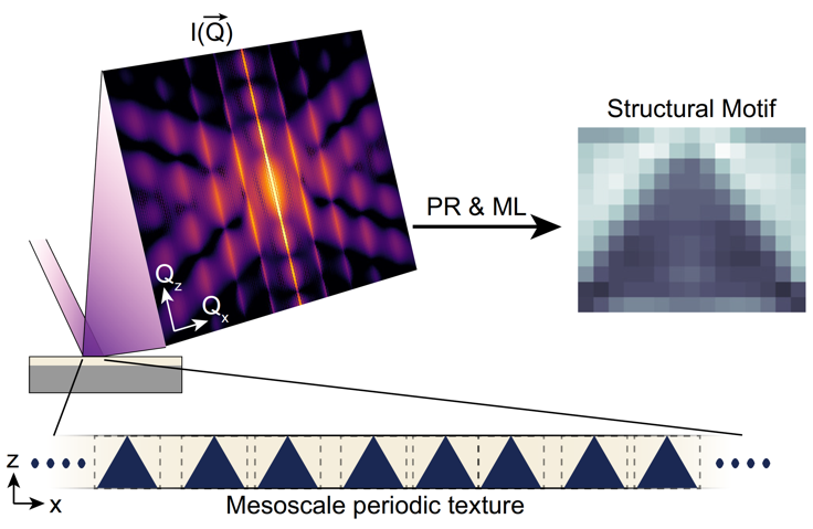
Figure 1:
Concept of work. In a reciprocal space map, the mesoscale periodic ordering is evident from satellite peaks around the Bragg reflection. The diffraction intensity shown originates from the schematic periodic textures with triangular motifs, where the dark areas have a constant strain difference compared to the surrounding light area.
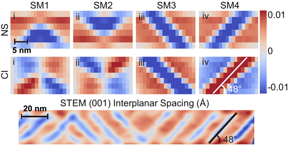
Figure 2: X-ray imaging (top two rows): Nanotextures calculated to be present in a strained Ca2RuO4 film measured at 7 K. (A) Measured and (B) reconstructed diffraction pattern. Four characteristic structural motifs (SMs) labelled normal strain (NS) and crystal-plane inclination (CI) are identified.
Electron Microscopy (bottom): Cryo-STEM map of the interplanar spacing along [001] of the cross-section of a ~34-nm-thick Ca2RuO4 film at ~100 K. The features seen in STEM corroborate the nanotextures calculated from X-ray diffraction data, including the average stripe angle (48°).
The method solves a long-standing problem and achieves a direct, model-independent inversion of x-ray diffraction data. Using the technique, a previously unreported nanotexture in a film undergoing a metal-insulator transition (Ca2RuO4) is discovered and confirmed by PARADIM’s cryo-STEM capabilities (Figure 2). The method is relevant to modulated structures in ferroics and topological quantum materials.
New properties and exotic quantum phenomena can form due to periodic nanotextures, including moiré patterns, ferroic domains, and topologically protected magnetization and polarization textures. Despite the availability of powerful tools to characterize the atomic crystal structure, the visualization of nanoscale strain-modulated structural motifs remains challenging. Here, nondestructive real-space imaging of periodic lattice distortions in thin epitaxial films is demonstrated, showing an emergent periodic nanotexture in a Mott insulator. Specifically, the approach combines iterative phase retrieval with unsupervised machine learning to invert the diffuse scattering pattern from conventional X-ray reciprocal-space maps into real-space images of crystalline displacements. The imaging in PbTiO3/SrTiO3 superlattices exhibiting checkerboard strain modulation substantiates published phase-field model calculations.
Furthermore, the imaging of biaxially strained Mott insulator Ca2RuO4 reveals a strain-induced nanotexture comprised of nanometer-thin metallic-structure wires separated by nanometer-thin Mott-insulating-structure walls, as confirmed by cryogenic scanning transmission electron microscopy (cryo-STEM). The nanotexture in Ca2RuO4 film is induced by the metal-to-insulator transition and has not been reported in bulk crystals. One can expect that the phasing of diffuse X-ray scattering from thin crystalline films in combination with cryo-STEM will open a powerful avenue for discovering, visualizing, and quantifying the periodic strain-modulated structures in quantum materials.
Coherent X-ray diffractive imaging (CXDI) has been widely applied to study the strain distribution in confined systems using an illuminating beam with high spatial coherence. Here, we broaden the capability of CXDI by imaging the strained structural motifs in periodic nanotextured epitaxial thin films with unprecedented nanoscale resolution, exploiting the diffuse scattering pattern from a conventional, partially coherent synchrotron beam. We demonstrate the technique by discovering a previously unreported nanotexture in a Mott insulator epitaxial thin film, which is confirmed by atomic resolution cryo-STEM imaging. The work promises to become a stepping stone for quantifying periodic strain-modulated structures in ferroics and topological quantum materials.
PARADIM’s Electron Microscopy Facility enabled direct visualization of the novel nanotexture at atomic resolution with cryogenic scanning transmission electron microscopy, complementing and contributing to the development of the new x-ray technique.
The work was initialized by users from Cornell University and completed in collaboration with researchers at Penn State University, Cornell High-Energy Synchrotron Source (CHESS), and the Advanced Photon Source (APS).
Z. Shao, N. Schnitzer, J. Ruf, O.Y. Gorobtsov, C. Dai, B.H. Goodge, T. Yang, H. Nair, V.A. Stoica, J.W. Freeland, J. Ruff, L.-Q. Chen, D.G. Schlom, K.M. Shen, L.F. Kourkoutis, and A. Singer, "Real-Space Imaging of Periodic Nanotextures in Thin Films via Phasing of Diffraction Data," Proc. Natl. Acad. Sci. U.S.A. 120, e2303312120 (2023). DOI: 10.1073/pnas.2303312120
The work was primarily supported by U.S. Department of Energy, Office of Science, Office of Basic Energy Sciences, under contract no. DE-SC0019414 (development of the phase retrieval algorithm, data analysis, and interpretation: Z.S., O.Y.G., D.G.S., K.M.S., A.S.; thin-film synthesis: H.N.). This research was funded in part by the Gordon and Betty Moore Foundation’s EPiQS Initiative through grant nos. GBMF3850 and GBMF9073 to Cornell University. Sample preparation was, in part, facilitated by the Cornell NanoScale Facility, a member of the National Nanotechnology Coordinated Infrastructure (NNCI), which is supported by the NSF (grant no. NNCI-2025233). We thank Benjamin Gregory for careful reading of the manuscript. Research conducted at CHESS was supported by the NSF (BIO, ENG and MPS Directorates) under awards DMR-1332208 and DMR-1829070. Use of the Advanced Photon Source was supported by DOE’s Office of Science under contract DE-AC02-06CH11357. Transmission electron microscopy work was supported by the NSF [Platform for the Accelerated Realization, Analysis, and Discovery of Interface Materials (PARADIM)] under Cooperative Agreement No. DMR-2039380 and made use of the Cornell Center for Materials Research Shared Facilities, which are supported through the NSF MRSEC program (DMR-1719875). N.S. was supported by the NSF Graduate Research Fellowship (DGE-2139899). B.H.G. was supported by PARADIM (NSF DMR-2039380). L.F.K. acknowledges support by the Packard Foundation. T.Y. and L.-Q.C.’s efforts on phase-field simulations are supported as part of the Computational Materials Sciences Program funded by the U.S. Department of Energy, Office of Science, Basic Energy Sciences, under award no. DE-SC0020145. C.D.’s effort is supported by the U.S. Department of Energy, Office of Science, Office of Basic Energy Sciences, under award number DE-SC-0012375. V.A.S. and J.W.F. acknowledge the U.S. Department of Energy, Office of Science, Office of Basic Energy Sciences, under award number DE-SC-0012375, for support in studying complex-oxide heterostructure with X-ray scattering.